How it works
Ultrastructural analysis of podocytes
Podocyte Exact Morphology Measurement Procedure – PEMP is a quantitative and quick procedure measuring the slit membrane density. It is ideal for determining the exact foot process morphology in biopsies and kidney sections.
- Works with FFPE tissue from mice, rats, pigs, NHP, humans, and organoids
- Quantitative readouts:
- FSD (filtration slit density): Indicates the degree of effacement
- FSL (filtration slit length): Indicates the filtration potential of a glomerulus
PEMP is currently for research use only.
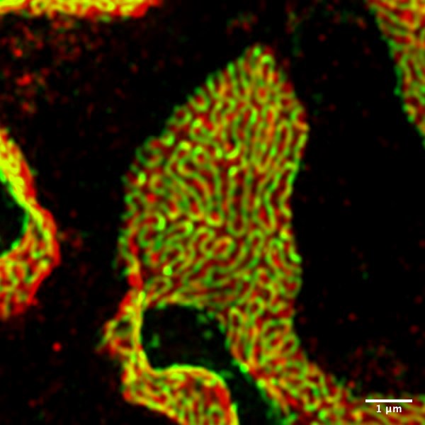
3D-Structured Illumination Microscopy (3D-SIM)
Podocyte foot processes have a size of only 100-200 nm. Until recently, the foot process could only be visualized using (transmission) electron microscopy. Now with the aid of the super-resolution microscopy, it is possible to make the podocytes’ foot processes and the filtration slit visible under a fluorescence light microscope.
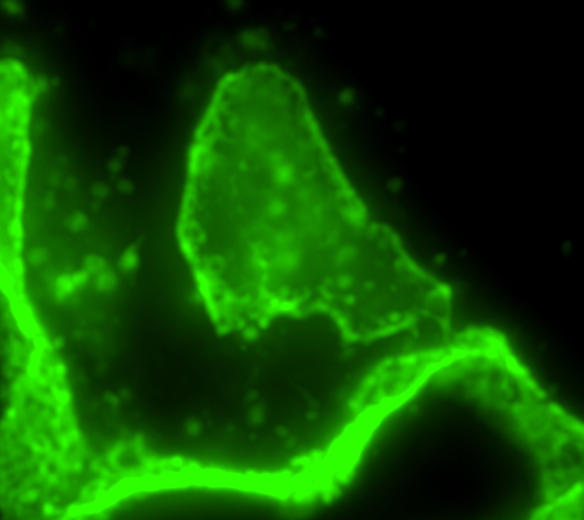
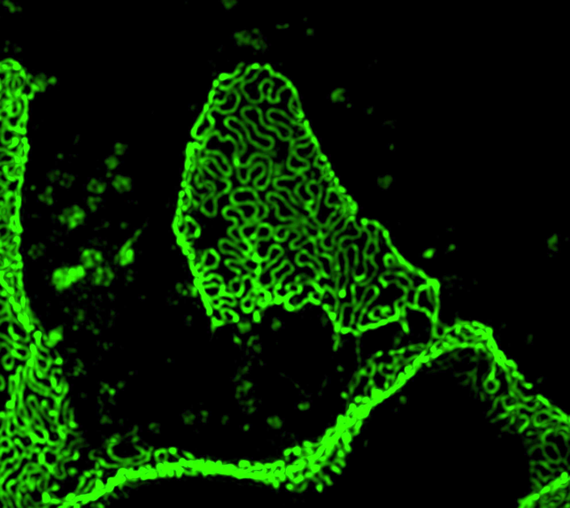
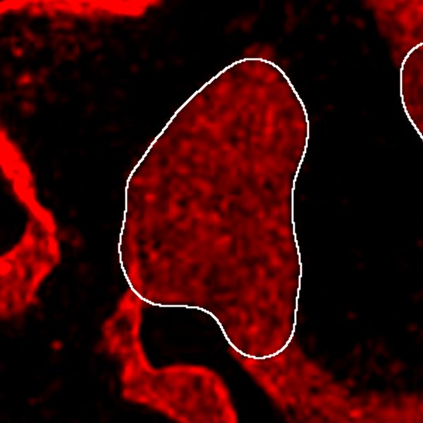
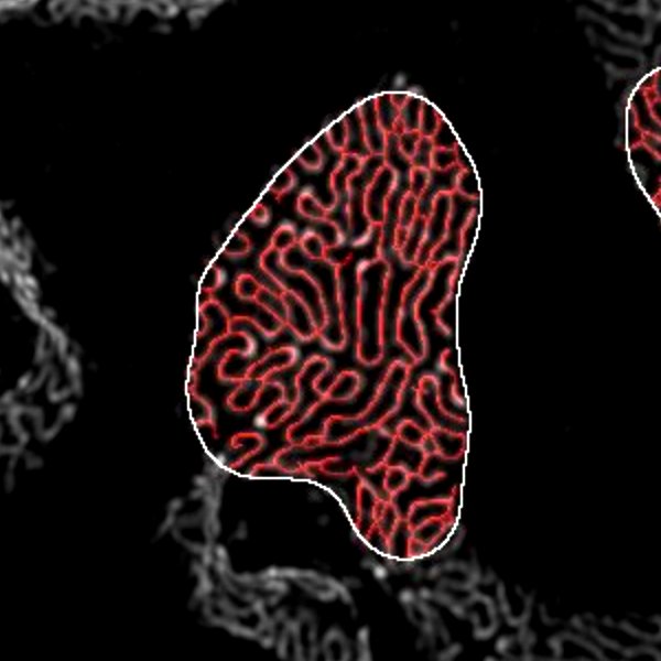
Kidney sample of a mice with Widefield Microscopy compared to 3D-SIM
Workflow
1. Preprocessing
Embedding, sectioning, and immunofluorescence staining using antibodies against podocin/nephrin and integrin/synaptopodin.
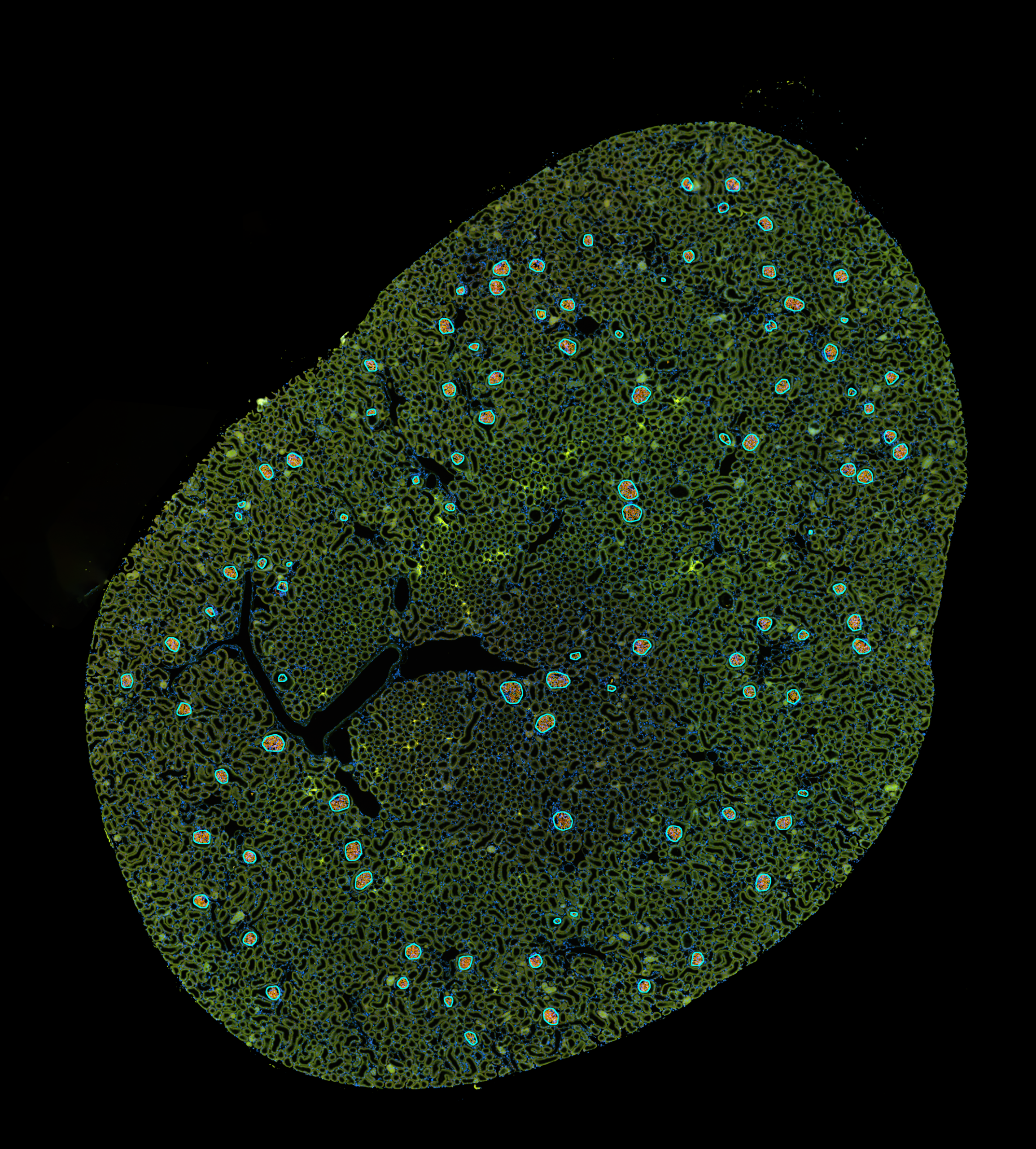
2. Whole Slide Imaging
10x whole-slide epifluorescence image
AI-based glomeruli segmentation and selection of glomeruli for super-resolution

3. Acquisition of 3D-SIM
Imaging of dual-channel z-stack of selected glomeruli and reconstruction into 3D-SIM image and maximum intensity projection (MIP).
4. Evaluation
Fully automated evaluation by identifying podocyte areas and measuring the filtration slit length.
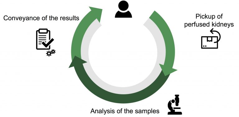
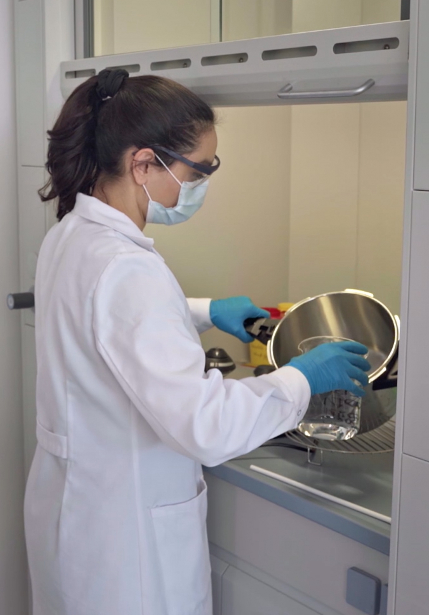
Our service – Send-and-forget.
Send us your samples and we will take care of everything else.