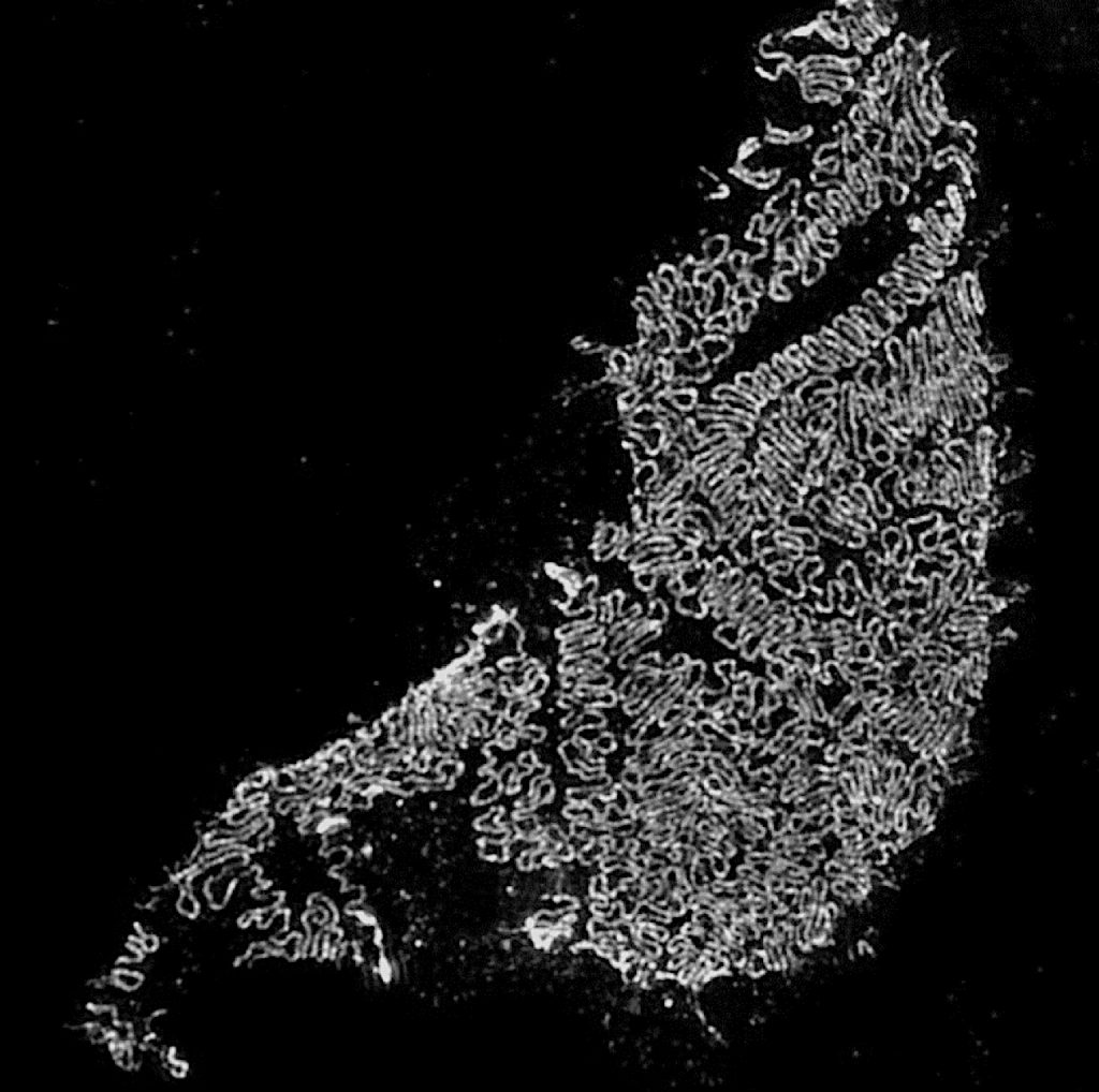
Quantify Podocyte Foot Process Morphology
We developed a new procedure based on Super-Resolution Microscopy that allows quick and precise quantification of podocyte foot process morphology.
How it works
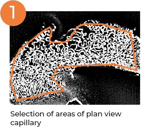
Selection
Selection of areas of plan view capillary
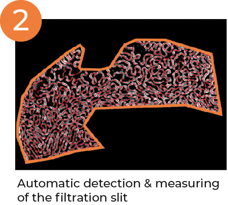
Identify
Automatic detection & measuring of the filtration slit
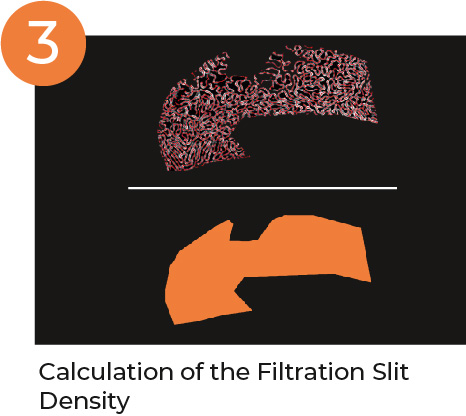
Analyze
FSD (Filtration Slit Density) Ratio of filtration slit length and selected area
Benefits
Single, objective & comparable value
Measurement per Glomerulus
Identification of small changes
High accuracy – Analysis of 12 to 20 Glomeruli per microscopic slice
Staining with up to 3 antibodies (WT1, Synaptopodin, etc.)
PEMP in Peer-Reviewed Research
Structured illumination microscopy and automatized image processing as a rapid diagnostic tool for podocyte effacement.
Siegerist F, Ribback S, Dombrowski F, Amann K, Zimmermann U, Endlich K, Endlich N
Scientific Reports (2017) PMID 28904359
ARP3 controls the podocyte architecture at the kidney filtration barrier.
Schell C, Sabass B, Helmstaedter M, Geist F, Abed A, Yasuda-Yamahara M, Siglek A, Maier JI, Grahammer F, Siegerist F, Artelt N, Endlich N, Kerjaschki D, Arnold HH, Dengjel J, Rogg M, Huber TB
Developmental Cell (2018) PMID 30503751
Comparative analysis of podocyte foot process morphology in three species by 3D super-resolution microscopy
Artelt N, Siegerist F, Ritter AM, Grisk O, Schlüter R, Endlich K, Endlich N
Frontiers in Medicine (2018) PMID 30425988
Novel Microscopic Techniques for Podocyte Research
Siegerist F, Endlich K, Endlich N
Frontiers Endocrinol (2018) PMID 30050501
The Role of Palladin in Podocytes
Artelt N, Ludwig TA, Rogge H, Kavvadas P, Siegerist F, Blumenthal A, van den Brandt J, Otey CA, Bang ML, Amann K, Chadjichristos CE, Chatziantoniou C, Endlich K, Endlich N
Journal of the American Society of Nephrology (2018) PMID 29720549
Cosmc-dependent Mucin-Type O-linked Glycosylation Is Essential for Podocyte Function
Stotter BR, Talbot BE, Capen DE, Artelt N, Zeng J, Matsumoto Y, Endlich N, Cummings RD, Schlondorff JS
Am J Physiol Renal Physiol (2020) PMID 31904283
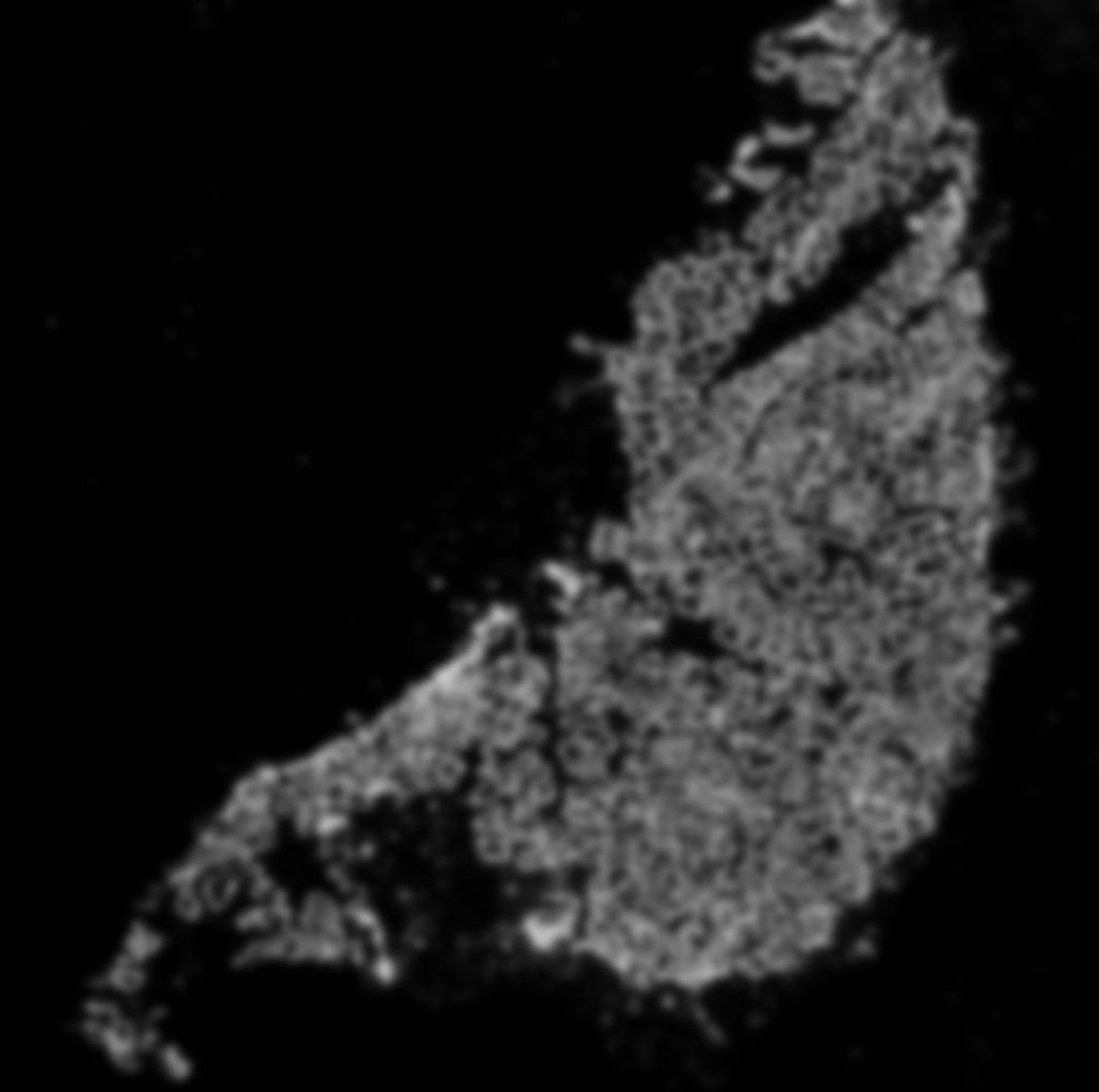
3D-Structured Illumination Microscopy
Podocyte foot processes have a size of only 100-200 nm. Until recently, these cells could only be visualized using the electron microscopy. Now with the aid of the super-resolution microscopy it is possible to make the podocytes foot processes and the filtration slit visible under a fluorescence light microscope.
Kidney sample of a mice with Widefield Microscopy compared to 3D-SIM